
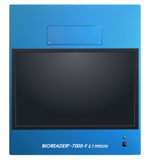
BIOREADER® 7000 -F-z-i micro
Versatile inverted Fluoro/VIS Reader, micro + macro zoom, high power LED for fluoro applications
The Bioreader® 7000 -F-z-i micro provides an automatic zoom range for full-field-of-view of 35 - 100 mm Petridish, 6 - 384 well plates and max. 1 x 1 mm zoom factor on microscopic slides or hemocytometer.
Optical resolution for good shape recognition: Recommended from 10 µm(1) for single cells, 10 - 2000 µm for FOCI, 100 - 5000 µm for plaques or 50 - 2000 µm for cell clones.
(1) Depending on the selected front lens on the micro zoom (2x, 3x or 4x).
Model variants:
Fluoro filtersets installed Max. VIS light possible
Alpha 1 3
Beta 2 3
Gamma 3 3
Delta 6 3
All Bioreader® 7000 -F-z-i micro model variants can be upgraded to max. 8 Fluoro filtersets.
Independence
Our products operate independent from any assay or kit manucafurer. So you may choose among all assay/kit manufactures.
Antiviral vaccine development by use of the Bioreader®
Our products support testing and development of antiviral vaccines.
Example#1: The automatic analyses of Plaque reductions assay.
This assay applies diluted doses of a -possibly- antiviral drug into microwell plates. Our products automatically observe its capability to suppress the formation of plaques (-holes-) in the bacterial lawn.
The ‘Plaque reductions neutralization test’ (PRNT) is a variation of this test. It is an attempt to find an antibody that neutralizes a specific virus. This is a very valuable tool in antiviral vaccine research.
Examplel#2: The automatically evaluation of the Yield reduction assay
In several steps of operation cell are infected in presence of variable doses of a possibly antiviral drug substance.
Thereafter the virus titer is determined in the liquid by use of the plaque or TCID50 assay by the use of our products.
The titer is calculated as the quotient of the positive cells/all cells, taking in account the virus dilution factor and the volume of the inoculum.
- Full automation of all mechanical functions incl. focus, zoom, aperture.
- 8 times filter changer.
- Upgrade option from model alpha to gamma and delta.
- Dual telecentric illumination for better cytokine quantification.
- Automated zoom lens for 6-48 well plates and/or Petri dishes.
- Fixed focus lens for 96-384 well plates or Petri dishes.
- Microscopic zoom system for microscopic cell counting.
- Fluoro/UV LED excitation plus 3 types of white light LED illuminations.
- Hardware is automatically controlled by software measure setup (GxP-locked).
- No mercury or deuterium lamps required, thus 50T hourly lifetime.
- Improved suppression of auto-fluorescence, cluster separation, well recognition and cytokine volume quantification.
- Brand-new generation 7 makes it significant faster and more precise regarding positioning.
- Single click export to PDF, Microsoft Office® applications.
- Front loader, automatic door.
- Dual tele-centric illumination.
- Loud speaker and interactive training program.
- Excel/Word/Power Point and LIMS export capabilities.
- Suitable for research purpose.
- High contrast/low noise cameras 5-41 MPixel available.
- Optional full automation with plate 'feeding system'.
- DQ/IQ/OQ/PQ documention possible.
- 12 months warranty.
- Declaration of Conformity and EMC certificate.

- Innovative Bioreader® software combines ‘easy of use’ and versatility and flexibility.
- Scan, analyze and overlay live time.
- Creates up to 7 images for each well simultaneously during the scan.
- ‘Profiling’ app helps to create user independent measure protocols for Elispot beginners and references for experts.
- Verified in collaborative studies.
- Export options and reports with all scalable images and results even in one file.
- Customer specific export and report templates.
- Video clips and content specific help files.
- Qualified installation and training with each Bioreader® model.
- On-site or internet remote services and support.
- ‘Classified’ measure protocols ‘history’ tracking and comparison tools.
- User specific plates, Studies/projects, plate layouts/designs and measure protocols prevent from mix-up.
- More accurate ‘cytokine quantification’ based on the patented ‘photometric’ dual illumination system.
- Optional software features:
- 21 CFR part 11 based software module available
Dimension
Weight
Cell compartment visualization and cell transfection efficiency
Cell compartment visualization and cell transfection efficiency
It is possible by use of the Bioreader® 7000-F-z with micro-resolution to determine the transfection rate. Differciation of compartments should be possible if compartments are transfected with specific proteins and labelled with individual fluorophores.
Elispot in 384 microfilter plates
Small volume, higher throughput.
Elispot - enzymatic single color
Elispot single encymatic
Blue, red, green or silver substrat Elispot.
Detection antibody, enzyme-conjugate and precipitating substrate.
"The enzyme-linked immune absorbent spot (ELISpot) is a type of assay that focuses on quantitatively measuring the frequency of cytokine secretion for a single cell."
[https://en.wikipedia.org/wiki/ELISpot]
Why to use Elispot
•The cytokine Elispot assay is designed to enumerate cytokine secreting cells in single cell suspensions of lymphoid tissue, CNS tissue, bone marrow or preparations of peripheral blood mononuclear cells (PBMC).
•The assay has the advantage of detecting only activated/memory T cells and the cytokine release can be detected at the single cell level, allowing direct determination of T cell frequencies.
•The assay has the advantage of detecting only activated/memory T cells and the cytokine release can be detected at the single cell level, allowing direct determination of T cell frequencies.
The ELISPOT assay is an effective tool to enumerate antigen-specific T cells in the circulation of immunized humans and animals at much lower frequencies than possible with other currently available methods
The ELISPOT assay has proven to be a sensitive and unique system to follow disease progression in human individuals or animals. Several studies have indicated that alterations in the frequency of cytokine pc in different compartments of the body adequately reflect changes in immune function
The ELISPOT assay may be used to determine effects of drugs, chemicals or other compounds on cytokine secretion in vitro, thereby providing data on their putative modulatory effects on immune function in vivo
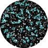
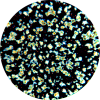
Fluorospot 1-4 fluorophores
Multi Fluoro Color Reader,
Best for Elispot and FluoroSpot.
Applies ‘local neighborhood processing’ in order to compensate for uneven background on fluoro plates. This way the spot volume is quantified, independent of the local background.
Why use Fluorospot?
Enzymatic Elispot labeling
• allows to label 1-2 cytokines per cell simultanously.
• If you are interested to see ‘double secreting’ cells the stain of two cells in neighborhood may overlap. So, It is not clear if two cells are overlapping or one cells is really secreting two different cytokines ( see below left)
‘Fluorospot’ labeling
Allows to label -a theoretical unlimited number different- cytokines. Because excitation for each cytokine is applied -one after the other-, decisions for double secreting cells is clearly.
One Image is acquired for each cytokine!
Currently Fluorospot assays with 1-4 Fluorophores are on the market.
Fluorospot offer's significant advantages over colorimetric formats, particularly in the areas of multiplexing and automated spot detection.
Moreover, as spot development is not enzymatic, signal intensity is directly proportional to the amount of analyte within.
Infection assay
Cell infection assay
Plaque reduction assay and Yield reduction assay are approved methods to determine antiviral substances
Verify virus titer by use of TCID50 (Median Tissue Culture Infectious Dose)
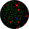
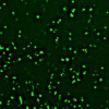
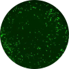
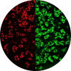
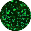
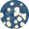
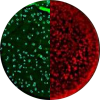
Example: Green fluorescence. Adherent cells (primary human fibroblasts) in 96-well-mikrotiterplate. Fluorophores: DAPI and AlexaFluor488.
Microscopic magnification for immunohistochemical and fluorophores staining for plates from 6 to 384 wells and Microscope/Hemocytometer slides.
>
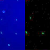
GFP/DAPI-expressed virus infected
GFP/DAPI-expressed virus infected
"Using DNA recombinant technology, scientists combine the GFP gene to a another gene that produces a protein that they want to study, and then they insert the complex into a cell." [https://embryo.asu.edu/pages/green-fluorescent-protein]
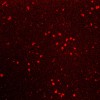
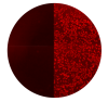
RFP-expressed virus infected
RFP-expressed virus infected
"Using DNA recombinant technology, scientists combine the RFP gene to a another gene that produces a protein that they want to study, and then they insert the complex into a cell." [https://embryo.asu.edu/pages/green-fluorescent-protein but with RFP instead of GFP]
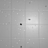
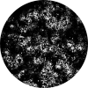
non stained cells
Non stained cells may be counted in a kind of phase contrast mode.
Alveolar epithelial cells (AEC)
Alveolar epithelial cells (AEC)
"Typically, type 1 alveolar cells comprise the major gas exchange surface of the alveolus and are integral to the maintenance of the permeability barrier function of the alveolar membrane. Type 2 pneumocytesare the progenitors of type 1 cells and are responsible for surfactant production and homeostasis." [https://www.sciencedirect.com/topics/medicine-and-dentistry/alveolar-type-i-cells]
The alveolar epitheliumis a major target in toxic exposures of the lung because of its structural delicacy and proximity to inhaled toxicants. Type II epithelial cellsare important in maintaining the integrity of alveolar epitheliumand normal lung function.
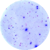
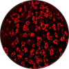
Elispot - enzymatic dual color
Blue and red Elispot substrate (mixed color violet)
Dual cytokine secretion.
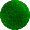
Cell viability
Cell viability
6-CF appears as green fluorescence.
Live/dead
Live/dead cells
Live cells are white, dead cells are black. Tryphaneblue stain.
Transgene assay
Transgene assay
"In its most precise usage, the term transgenedescribes a segment of DNA containing a gene sequence that has been isolated from one organism and is introduced into a different organism." [https://en.wikipedia.org/wiki/Transgene]
Cells transfected with an expression plasmid encoding eGFP.
Live single cells
Live single cells
Bright field application Jurkatcells.
Live/apoptotic cells
Live/apoptotic cells
NUCblue/interference/NUCgreen. Mix of Jurkatcells untreated cell proliferate and TNF/CHX-treated that causes to apoptosis.
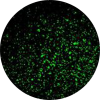
MCMV (Murine Cytomegalovirus) greenfluoro remission
MCMV (Murine Cytomegalovirus)
green fluoro remission
"Cytomegalovirus (CMV) (from the Greek cyto-, "cell," and megalo-, "large") is a genus of viruses in the order Herpesvirales." [https://en.wikipedia.org/wiki/Cytomegalovirus]
MCMV (Murine Cytomegalovirus) red fluoro remission.
MCMV (Murine Cytomegalovirus)
red fluoro remission
MCMV: mouse cytomegalovirus, unstained
MCMV: mouse cytomegalovirus, unstained
HCMV Human cytomegalovirus, unstained
HCMV human cytomegalovirus, unstained
"The glycoprotein gO (UL74) of human cytomegalovirus (HCMV) forms a complex with gH/gL. Virus mutants with a deletion of gO show a defect in secondary envelopment with the consequence that virus spread is restricted to a cell-associated pathway." [https://www.ncbi.nlm.nih.gov/pubmed/20181688]
MCMV mutants got generated that express with tdTomato, eGFP or mCherry
MCMV mutants got generated that express with tdTomato, eGFP or mCherry
MCMV mutants, tdTomato, Cherry, eGFP fibroplasts
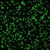
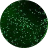
NK92 cells natural killer cells (human cell line) expressing green fluorescent protein (gfp)
NK92 cells natural killer cells (human cell line) expressing green fluorescent protein (gfp)
"Natural killer cells, or NK cells, are a type of cytotoxic lymphocyte critical to the innate immune system. Typically, immune cells detect major histocompatibility complex (MHC) presented on infected cell surfaces, triggering cytokine release, causing lysis or apoptosis." [https://en.wikipedia.org/wiki/Natural_killer_cell]
GFP immunofluorescence on HEK 293 cells
GFP immunofluorescence on HEK 293 cells
"Human embryonic kidney 293 cells, also often referred to as HEK 293, HEK-293, 293 cells, or less precisely as HEK cells, are a specific cell line originally derived from human embryonic kidney cellsgrown in tissue culture." [https://en.wikipedia.org/wiki/HEK_293_cells]
NK92 cells natural killer cells (human cell line) same samples microwell plate.
NK92 cells natural killer cells (human cell line) same samples microwellplate
"Natural killer cells, or NK cells, are a type of cytotoxic lymphocyte critical to the innate immune system. Typically, immune cells detect major histocompatibility complex (MHC) presented on infected cell surfaces, triggering cytokine release, causing lysis or apoptosis." [https://en.wikipedia.org/wiki/Natural_killer_cell]
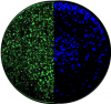
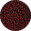
Mouse spleen cells mostly lymphocytes stained with FITC-conjugated anti mouse cd45 stain
Mouse spleen cells mostly lymphocytes stained with FITC-conjugated anti mouse CD45 stain
"The spleen is an organ found in virtually all vertebrates. Similar in structure to a large lymph node, it acts primarily as a blood filter. A study published in 2009 using mice found that the red pulp of the spleen forms a reservoir that contains over half of the body's monocytes." [https://en.wikipedia.org/wiki/Spleen]
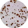
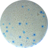
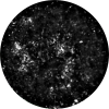
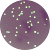
Foci assay: total well view
Foci assay: total well view
"The focus forming assay (FFA) is a variation of the plaque assay, but instead of relying on cell lysis in order to detect plaque formation, the FFA employs immunostaining techniques using fluorescently labeled antibodies specific for a viral antigen to detect infected host cells and infectious virus particles before an actual plaque is formed." [https://en.wikipedia.org/wiki/Virus_quantification]
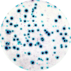
Foci assay: Very large Foci
Foci assay: very large Foci
"The focus forming assay (FFA) is a variation of the plaque assay, but instead of relying on cell lysis in order to detect plaque formation, the FFA employs immunostaining techniques using fluorescently labeled antibodies specific for a viral antigen to detect infected host cells and infectious virus particles before an actual plaque is formed." [https://en.wikipedia.org/wiki/Virus_quantification]

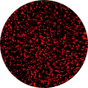
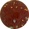
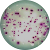
Plaque assay extremely large spots
Plaque assay with extremely large spots
"The TCID50 (Median Tissue Culture Infectious Dose) is one of the methods used when verifying viral titer. TCID50 signifies the concentration at which 50% of the cells are infected when a test tube or well plate upon which cells have been cultured is inoculated with a diluted solution of viral fluid." [https://www.zeomic.co.jp/en/glossary/virus/71]
TCID50 viral assay
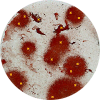
CV Plaque assay.Example with large and fuzzy plaques
CV Plaque assay; example with large and fuzzy plaques
Cells were infected and covered with an overlay. Surviving cells stained with CV. Cells killed by the virus.

Plaque assay.Very bright plaques in 24 well plate
Plaque assay with very bright plaques in 24 well plate
Cells were infected and covered with an overlay. Surviving cells stained with CV. Cells killed by the virus.
Neutralization assay
Neutralization assay
"The plaque reduction neutralization test is used to quantify the titer of neutralizing antibody for a virus. The serum sample or solution of antibody to be tested is diluted and mixed with a viral suspension." [https://en.wikipedia.org/wiki/Plaque_reduction_neutralization_test]
Neutralization assay Accepted Plaques are surrounded
Neutralization assay
Accepted Plaques are surrounded
"The plaque reduction neutralization test is used to quantify the titer of neutralizing antibody for a virus. The serum sample or solution of antibody to be tested is diluted and mixed with a viral suspension." [https://en.wikipedia.org/wiki/Plaque_reduction_neutralization_test]
Detection of Marek's disease virus Plaques after staining with specific anti-MDV antibodies and Alexa 488 labelled secondary antibodies
Detection of Marek's disease virus
Plaques after staining with specific anti-MDV antibodies and Alexa 488 labelled secondary antibodies
"Marek's disease is a highly contagious viral neoplastic disease in chickens. The disease is characterized by the presence of T cell lymphoma as well as infiltration of nerves and organs by lymphocytes. Viruses related to MDV appear to be benign and can be used as vaccine strains to prevent Marek's disease." [https://en.wikipedia.org/wiki/Marek's_disease]
Detection of Marek's disease virus Plaques after staining with specific anti-MDV antibodies and Alexa 568 labelled secondary antibodies
Detection of Marek's disease virus but Alexa 568 labelled
"Marek's disease is a highly contagious viral neoplastic disease in chickens. The disease is characterized by the presence of T cell lymphoma as well as infiltration of nerves and organs by lymphocytes. Viruses related to MDV appear to be benign and can be used as vaccine strains to prevent Marek's disease. " [https://en.wikipedia.org/wiki/Marek's_disease]
Organoids
Organoids
"An organoid is a miniaturized and simplified version of an organ produced in vitro in three dimensions that shows realistic micro-anatomy. The technique for growing organoids has rapidly improved since the early 2010s." [https://en.wikipedia.org/wiki/Organoid]
Dual Transgene assay
Dual Transgene assay
A segment of DNA introduced to some other organism. See [https://en.wikipedia.org/wiki/Transgene]
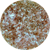
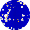
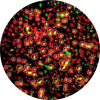
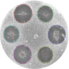
Various colonies in 6 well plates and Petridishes 35 mm - 100 mm
Various colonies in 6 well plates
Various media/strain combinations on petridishes
Various media/strain combinations on petridishes
Antibiotic potency USP/PhEur/DIN/BP Petrishes
The antibiotic potency assay measures the effectiveness of an antibiotic by the degree of growth inhibition on susceptible strains of microorganisms at different concentrations.
Adenovirus
Adenovirus
"Adenoviruses (members of the family Adenoviridae) are medium-sized (90–100 nm), nonenveloped (without an outer lipid bilayer) viruses with an icosahedral nucleocapsid containing a double stranded DNA genome. Their name derives from their initial isolation from human adenoids in 1953.[1] They have a broad range of vertebrate hosts; in humans, more than 50 distinct adenoviral serotypes have been found to cause a wide range of illnesses, from mild respiratory infections in young children (known as the common cold) to life-threatening multi-organ disease in people with a weakened immune system."[https://en.wikipedia.org/wiki/Adenoviridae]
SARS COV-2
Vero cells infected by SARS-CoV-2
Inverted Immune fluorescence.
Secondary Antibody Cy3 AffiniPure Goat Anti-Human IgG
Bioreader® 7000 -F-Z-i Micro
The versatile multiwell plate evaluation system for testing antiviral substances against SARS-COV2, HDV, hepatitis and other virus groups.
Many laboratories are currently researching the mechanisms by which viruses enter cells (with a focus on SARS-CoV-2).
Various proteins on the surface of the cells play a major role here. These are a present in different amounts from test person to test person.
The currently occurring coronavirus SARS-CoV-2 has the capability to penetrate the cell by the help of even small amounts of this specific proteins. Extensive research is required to develop substances that reduce or prevent the virus from entering the cell.
This is where the Bioreader® 7000 -F-z-i Micro comes into play, which allows the researcher to test a large number of antiviral substances in vitro. The Bioreader® is currently used in leading national and international virological research laboratories with a large number of different cell lines and different viruses.
Another established method is the end-point limiting dilution assay for determining the mean Tissue Culture Infectious Dose (TCID50), which allows a statement to be made about how much the infection of the virus is slowed down or prevented by a substance.
In these and other in vitro tests, it is necessary to use extremely variable optical magnification so that plaques, cell clusters, foci as well as single cells are well imaged.
The cells / plaques / foci etc. must be countable from Petri dishes, cell culture bottles, 6-384 well plates and ultimately also microscope slides, both in white light and with various fluoro stimuli. With the Bioreader® 7000 -F-Z-i Micro the entire well /disk can be counted in full field of view or true optically enlarged partial areas.
The Bioreader® 7000 -F-z-i Micro is ideally suited for these tasks and is already used in many laboratories for research into antiviral substances.
The Bioreader® 7000 -F-z-i Micro is characterized by the fact that it has three separate optical systems which are used automatically depending on the format used (Petri dishes / cell culture bottles, 6-384 multi-well plates, microscope slides). All measurement parameters (with over 100 setting parameters) are applied automatically, especially for the respective application.
Here, e.g. a differentiation between infected and non-infected single cells can be made.