


BIOREADER® 7000 -F
Fluoro Elispot/Plaque + Enzymatic Readers for 96 wells with innovative power LED fluoro excitation.

This BIOREADER® 7000 -F allows you to evaluate verified enzymatic Elispot/Plaque or Fluorospot counting.
They are equipped with a long-lasting LED fluorescent light source. This allows you to evaluate verified enzymatic Elispot or Fluorospot counting.
The BIOREADER® 7000 -F can automatically apply up to 10 standard Olympus® fluoro filter cubes.
Model variants:
Fluoro filter cubes installiert Max. filter cubes possible
Alpha 2 6
Beta 3 6
Gamma 4 10
Delta 6 10
The Delta model has an additonal UV LED light source installed for DAPI filters.
The filter changer of the Bioreader® 7000 -F may be upgraded on site. Brickstone principle.
Independence
Bioreader® and EazyReader® operate with all commonly used Elispot/Fluorospot/Plaque/Fluoro-Plaque/CFU assays.
You can choose among all major assay manufactures.
- 6- or 10-times fluoro filter changer: For Standard Olympus® filter cubes on all models.
- New high sensitive but low noise camera technology with optimal lens glasses for high contrasted images.
- Filter upgrade options: From model Alpha up to Delta (max 10 filter cubes).
- 3D spot-volume cytokine quantification: Provides more sensitive stimulation response results.
- Bioreader® 7000 -F is an affordable model for fluoro and enzymatic Elispot and Plaque/FOCI counting in 384/96 well plates.
- Dual tele-centric illumination.
- Counting significantly faster and more precise regarding positioning.
- Front loader, automatic door.
- Loud speaker and interactive training program.
- Excel/Word/Power Point and LIMS export capabilities.
- Suiteable for reserarch purpose.
- Camera upgrade to 6 - 41 MPixel available.
- Optional automation with plate 'feeding system'.
- DQ/IQ/OQ/PQ documentation possible
- 12 months warranty.
- EC declaration of conformity and EMC certificate.

- Innovative Bioreader® and EazyReader® software combines ‘easy of use’ and versatility and flexibility.
- Scan, analyze and overlay live time.
- Creates up to 7 images for each well simultaneously during the scan.
- ‘Profiling’ app helps to create user independent measure protocols for Elispot beginners and references for experts.
- Verified in collaborative studies.
- Export options and reports with all scalable images and results even in one file.
- Customer specific export and report templates.
- Video clips and content specific help files.
- Qualified installation and training with each instrument.
- On-site or internet remote services and support.
- ‘Classified’ measure protocols ‘history’ tracking and comparison tools.
- User specific plates, Studies/projects, plate layouts/designs and measure protocols prevent from mix-up.
- More accurate ‘cytokine quantification’ based on the patented ‘photometric’ dual illumination system.
- Optional software features:
- 'Routine’ software package: Ooptimized applications for commonly used operations, only presents the icons you require for the specific job, quickly read a plate and quickly review, Q.C. and release the results.
- 21 CFR part 11 based software module available.
Dimension
Weight

Fluorospot 1-4 colors
Multi Fluoro Color Reader,
best for Elispot and FluoroSpot
Applies ‘local neighborhood processing’ in order to compensate for uneven background on fluoro plates. This way the spot volume is quantified, independent from the local background.
Why use Fluorospot?
Enzymatic Elispot labeling
• allows to label 1-2 cytokines per cell simultanously.
• If you are interested to see ‘double secreting’ cells the stain of two cells in neighborhood may overlap. So, It is not clear if two cells are overlapping or one cells is really secreting two different cytokines (see below left).
‘Fluorospot’ labeling
Allows to label -a theoretical unlimited number different- cytokines. Because excitation for each cytokine is applied -one after the other-, decisions for double secreting cells is clearly.
One Image is acquired for each cytokine!
Currently Fluorospot assays with 1-4 Fluorophores are on the market.
Fluorospot offer's significant advantages over colorimetric formats, particularly in the areas of multiplexing and automated spot detection.
Moreover, as spot development is not enzymatic, signal intensity is directly proportional to the amount of analyte within.
Elispot in 384 well microfilter plates
Small volume, higher throughput.
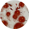
Elispot - Enzymatic single color
Blue, red, green or silver substrat Elispot.
Detection of antibodies. Enzyme-conjugated and precipitating substrate.
"The enzyme-linked immune absorbent spot (ELISpot) is a type of assay that focuses on quantitatively measuring the frequency of cytokine secretion for a single cell."
[https://en.wikipedia.org/wiki/ELISpot]
Why Elispot?
The cytokine Elispot assay is designed to enumerate cytokine secreting cells in single cell suspensions of lymphoid tissue, CNS tissue, bone marrow or preparations of peripheral blood mononuclear cells (PBMC).
The assay has the advantage of detecting only activated/memory T cells and the cytokine release can be detected at the single cell level, allowing direct determination of T cell frequencies.
The ELISPOT assay is an effective tool to enumerate antigen-specific T cells in the circulation of immunized humans and animals at much lower frequencies than possible with other currently available methods.
The ELISPOT assay has proven to be a sensitive and unique system to follow disease progression in human individuals or animals. Several studies have indicated that alterations in the frequency of cytokine pc in different compartments of the body adequately reflect changes in immune function.
The ELISPOT assay may be used to determine effects of drugs, chemicals or other compounds on cytokine secretion in vitro, thereby providing data on their putative modulatory effects on immune function in vivo
The ELISPOT assay is currently being used increasingly for the quantitative assessment of peptide reactive T lymphocytes from PBMC in infectious disease (3, 9) or in the course of vaccination trials aimed at the induction of tumor-specific T cells in patients with cancer.
Elispot may be used for Diagnosis of genetic defects,Allergic diseases,Autoimmune diseases,Transplantation,Cancer research,Acute inflammation,Acute infectious diseases and septic shock.
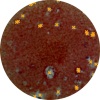
Elispot - Enzymatic dual color
Blue and red Elispot substrate (mixed color violet)
Dual cytokine secretion.
RFP-expressed virus infected
"Using DNA recombinant technology, scientists combine the GFP gene to a another gene that produces a protein that they want to study, and then they insert the complex into a cell."
[https://embryo.asu.edu/pages/green-fluorescent-protein but with RFP instead of GFP]
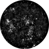
Unstained cells
Alveolar epithelial cells (AEC)
"Typically, type 1 alveolar cells comprise the major gas exchange surface of the alveolus and are integral to the maintenance of the permeability barrier function of the alveolar membrane. Type 2 pneumocytesare the progenitors of type 1 cells and are responsible for surfactant production and homeostasis."
[https://www.sciencedirect.com/topics/medicine-and-dentistry/alveolar-type-i-cells]
The alveolar epithelium is a major target in toxic exposures of the lung because of its structural delicacy and proximity to inhaled toxicants. Type II epithelial cells are important in maintaining the integrity of alveolar epithelium and normal lung function.

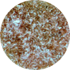
AEC graylevels
"Typically, type 1 alveolar cells comprise the major gas exchange surface of the alveolus and are integral to the maintenance of the permeability barrier function of the alveolar membrane. Type 2 pneumocytesare the progenitors of type 1 cells and are responsible for surfactant production and homeostasis."
[https://www.sciencedirect.com/topics/medicine-and-dentistry/alveolar-type-i-cells]
The alveolar epithelium is a major target in toxic exposures of the lung because of its structural delicacy and proximity to inhaled toxicants. Type II epithelial cells are important in maintaining the integrity of alveolar epithelium and normal lung function.
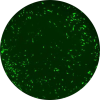
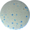
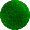
MCMV (Murine Cytomegalovirus) greenfluoro remission
"Cytomegalovirus (CMV) (from the Greek cyto-, "cell," and megalo-, "large") is a genus of viruses in the order Herpesvirales."
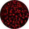
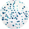
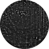
MCMV (Murine Cytomegalovirus) red fluoro remission.
"Cytomegalovirus (CMV) (from the Greek cyto-, "cell," and megalo-, "large") is a genus of viruses in the order Herpesvirales."
MCMV (Mouse Cytomegalovirus) unstained
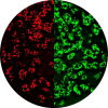
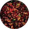
MCMV mutants got generated that express with tdTomato, eGFP or mCherry
MCMV mutants, tdTomato, Cherry, eGFP fibroplasts
HCMV (Human cytomegalovirus): unstained
"The glycoprotein gO (UL74) of human cytomegalovirus (HCMV) forms a complex with gH/gL. Virus mutants with a deletion of gO show a defect in secondary envelopment with the consequence that virus spread is restricted to a cell-associated pathway."
GFP immunofluorescence on HEK 293 cells
"Human embryonic kidney 293 cells, also often referred to as HEK 293, HEK-293, 293 cells, are a specific cell line originally derived from human embryonic kidney cells grown in tissue culture."
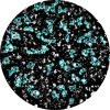
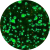
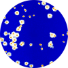
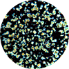
NK92 cells natural killer cells (human cell line) expressing green fluorescent protein (gfp)
"Natural killer cells, or NK cells, are a type of cytotoxic lymphocyte critical to the innate immune system. Typically, immune cells detect major histocompatibility complex (MHC) presented on infected cell surfaces, triggering cytokine release, causing lysis or apoptosis."
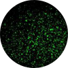
NK92 cells natural killer cells (human cell line) same samples microwell plate.
"Natural killer cells, or NK cells, are a type of cytotoxic lymphocyte critical to the innate immune system. Typically, immune cells detect major histocompatibility complex (MHC) presented on infected cell surfaces, triggering cytokine release, causing lysis or apoptosis."
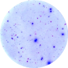
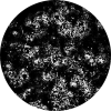
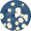
Mouse spleen cells mostly lymphocytes stained with FITC-conjugated anti mouse cd45 stain
"The spleen is an organ found in virtually all vertebrates. Similar in structure to a large lymph node, it acts primarily as a blood filter. A study published in 2009 using mice found that the red pulp of the spleen forms a reservoir that contains over half of the body's monocytes." [https://en.wikipedia.org/wiki/Spleen]
Foci assay: total well view
"The focus forming assay (FFA) is a variation of the plaque assay, but instead of relying on cell lysis in order to detect plaque formation, the FFA employs immunostaining techniques using fluorescently labeled antibodies specific for a viral antigen to detect infected host cells and infectious virus particles before an actual plaque is formed."
Foci assay: Very large Foci
"The focus forming assay (FFA) is a variation of the plaque assay, but instead of relying on cell lysis in order to detect plaque formation, the FFA employs immunostaining techniques using fluorescently labeled antibodies specific for a viral antigen to detect infected host cells and infectious virus particles before an actual plaque is formed."

GFP/DAPI-expressed virus infected
"Using DNA recombinant technology, scientists combine the GFP gene to a another gene that produces a protein that they want to study, and then they insert the complex into a cell."
Neutralization assay
"The plaque reduction neutralization test is used to quantify the titer of neutralizing antibodies for a virus. The serum sample or solution of antibodies to be tested is diluted and mixed with a viral suspension."
[https://en.wikipedia.org/wiki/Plaque_reduction_neutralization_test]
TCID50 viral assay
Cells were infected and covered with an overlay. Cells stained with CV survive. Unstained cells were killed by the virus.

Plaque assay extremely large spots
"The TCID50 (Median Tissue Culture Infectious Dose) is one of the methods used when verifying viral titer. TCID50 signifies the concentration at which 50% of the cells are infected when a test tube or well plate upon which cells have been cultured is inoculated with a diluted solution of viral fluid."
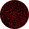
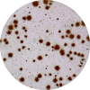
CV Plaque assay.Example with large and fuzzy plaques
Cells were infected and covered with an overlay. Cells stained with CV survive. Unstained cells were killed by the virus.

Plaque assay: very bright plaques in 24 well plate
Cells were infected and covered with an overlay. Cells stained with CV survive. Unstained cells were killed by the virus.
Organoids
"An organoid is a miniaturized and simplified version of an organ produced in vitro in three dimensions that shows realistic micro-anatomy. The technique for growing organoids has rapidly improved since the early 2010s."
Dual Transgene assay
Transgen:
A segment of DNA introduced to some other organism.